beginnings: V1 and grating orientations
Multiple groups, including Kamitani & Tong in their 2005 paper, suggest that this "hyperacuity" could be due to a voxel-level bias in the underlying architecture, whether in the distribution of orientation columns, the vasculature, or some combination of the two. The idea here is that, since columns are not perfectly evenly spatially distributed, each voxel will end up with more columns of one orientation than another, and this subtle population-level bias is what's being detected in the MVPA.
does degrading the signal reduce hyperacuity?
From the beginning, the idea that hyperacuity was behind the detection of orientation was met with both excitement and skepticism. In 2010 Hans op de Beeck wrote a NeuroImage Comments and Controversy article which kicked off a series of papers trying to confirm (or not) hyperacuity by means of degrading the fMRI images. The logic is straightforward: if subtle biases within individual voxels are making the classification possible, degrading the images (blurring, adding noise, filtering), should dramatically reduce the classification accuracy.op de Beeck (2010) smoothed images with information at varying spatial scales at different FWHM to change their signal, interpreting the results as suggesting that the apparent hyperacuity might actually be due to larger-scale patterns (spanning multiple voxels). Objections to this technique were raised, however, partly because smoothing's affect on information content is complex and difficult to interpret. Swisher et al. (2010) used spatial filters, rather than smoothing, to degrade the signal in very high-resolution images, and found that small (< 1 mm) scale information was present, and critical for classification accuracy. But, the presence of small-scale signal in high-resolution images doesn't preclude the presence of larger (> 2 mm) scale information; indeed, larger-scale information was also found by Swisher et al. (2010). Filtering was also used by Freeman et al. (2011), who identified larger ("coarser") scale information about orientation in V1. Alink et al. (2013) also used filtering, along with more complex stimuli, finding information in a range of scales, but also cautioning that the numerous interactions and complications mean that filtering is not a perfect approach.
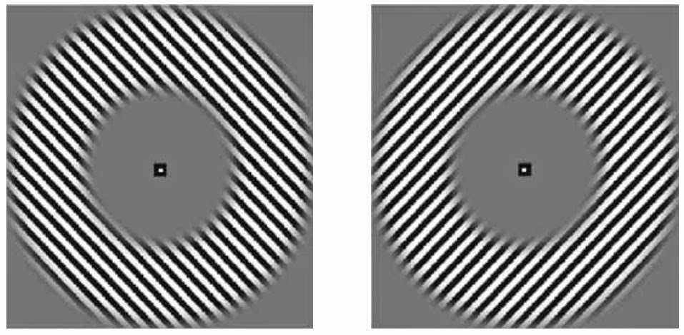
spiraling around

my musings, and does this imply anything about MVPA?
Overall, I'm landing in the "bigger patterns, not hyperacuity" camp. I find the demonstrations of larger-scale patterns convincing, and a more plausible explanation of the signal, at least for human fMRI with ~ 3 mm voxels; it strikes me as equally reasonable that very small-scale patterns could dominate for high-resolution scanning in anesthetized animals (e.g., Swisher et al. 2010).But do these debates imply anything for the usefulness of MVPA as a whole? Carlson and Wardle (2015) suggest that it does, pointing out that at this 10-year anniversary of the first papers suggesting the possibility of hyperacuity, we still haven't "determined the underlying source of information, despite our strong understanding of the physiology of early visual cortex." I wonder if this is because the best understanding of the physiology of early visual cortex is at the fine scale (neurons and columns), not the coarse scale ( > 5 mm maps). I agree that interpreting the properties of individual voxels from MVPA is fraught with difficulty; interpreting the properties of groups of voxels is much more robust.
papers mentioned here, plus some other relevant papers
- Alink, A., Krugliak, A.,Walther, A., and Kriegeskorte, N. (2013). "fMRI orientation decoding in V1 does not require global maps or globally coherent orientation stimuli." Frontiers in Psychology. doi:10.3389/fpsyg.2013.00493
- Beckett, A., et al. (2012) "Contribution of large scale biases in decoding of direction-of-motion from high-resolution fMRI data in human early visual cortex." NeuroImage. doi:10.1016/j.neuroimage.2012.07.066
- Carlson, TA. (2014). "Orientation Decoding in Human Visual Cortex: New Insights from an Unbiased Perspective." J Neuroscience. doi:10.1523/JNEUROSCI.0548-14.2014
- Carlson, TA and Wardle, SG. (2015). "Sensible Decoding." NeuroImage. doi:10.1016/j.neuroimage.2015.02.009
- Clifford, CWG and Mannion, DJ. (2015). "Orientation decoding: Sense in spirals?" NeuroImage. doi:10.1016/j.neuroimage.2014.12.055
- Freeman, J et al. (2011). "Orientation Decoding Depends on Maps, Not Columns" J Neuroscience. doi:10.1523/JNEUROSCI.5160-10.2011
- Freeman, J. Heeger, DJ,Merriam, EP. (2013). "Coarse-Scale Biases for Spirals and Orientation in Human Visual Cortex." J Neuroscience. doi: 10.1523/JNEUROSCI.0889-13.2013
- Haynes, JD. and G. Rees (2005). "Predicting the orientation of invisible stimuli from activity in human primary visual cortex." Nat Neuroscience. doi:10.1038/nn1445
- Kamitani, Y. and F. Tong (2005). "Decoding the visual and subjective contents of the human brain." Nat Neuroscience. doi:10.1038/nn1444
- Kriegeskorte, N., R. Cusack, et al. (2010). "How does an fMRI voxel sample the neuronal activity pattern: Compact-kernel or complex spatiotemporal filter?" NeuroImage. doi:10.1016/j.neuroimage.2009.09.059
- Kubilius, J. and Baeck, A. (2010) "From What Scale of Representation Does Multivariate Pattern Analysis Decode Information?" J Neuroscience. doi:10.1523/JNEUROSCI.1153-10.2010
- Gardner, J. (2010). "Is cortical vasculature functionally organized?" NeuroImage. doi:10.1016/j.neuroimage.2009.07.004
- Op de Beeck, H. P. (2010). "Against hyperacuity in brain reading: Spatial smoothing does not hurt multivariate fMRI analyses?" NeuroImage. doi:10.1016/j.neuroimage.2009.02.047
- Op de Beeck, H. P. (2010). "Probing the mysterious underpinnings of multi-voxel fMRI analyses." NeuroImage. doi:10.1016/j.neuroimage.2009.12.072
- Swisher, JD, et al. (2010). "Multiscale Pattern Analysis of Orientation-Selective Activity in the Primary Visual Cortex." J Neuroscience. doi:10.1523/JNEUROSCI.4811-09.2010
No comments:
Post a Comment